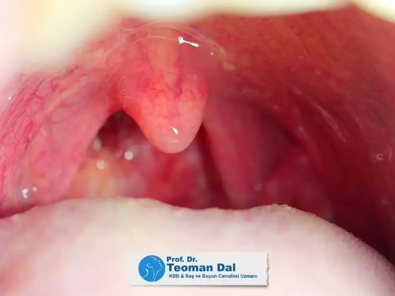
Obstrüktif uyku apnesi, uyku sırasında hava yolunun daralması veya tamamen tıkanması sonucu oluşan, ciddi sağlık sorunlarına yol açabilen bir durumdur. Bu tıkanıklık çoğu zaman birden fazla anatomik bölgede görülse de, dil kökü bölgesi en sık etkilenen yapılardan biridir. Uyku sırasında dili önde tutan kasların gevşemesiyle birlikte dil arkaya kayar ve bu da solunum yolunun daralmasına, horlamaya ve nefes durmalarına neden olur.
Özellikle orta ve ileri derecede uyku apnesi bulunan hastalarda dil kökü bölgesi tıkanıklığın ana kaynaklarından biri olarak karşımıza çıkar. Vücut kitle indeksi yüksek olan bireylerde, alt çenesi geride bulunan kişilerde ya da büyük dil hacmine sahip olan hastalarda bu sorun daha belirgindir. Bu hastalarda yalnızca yumuşak damak değil, dil kökü de solunumu engellediğinden tedavi planlaması buna göre yapılmalıdır.
Bu gibi vakalarda genellikle çok düzeyli hava yolu tıkanıklığı söz konusu olduğu için cerrahi planlama buna göre yapılmalıdır. Ameliyat öncesi:
- Kilo verme stratejileri
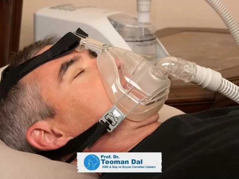
- CPAP cihazı kullanımı
- Uyku endoskopisi ile hava yolu değerlendirmesi mutlaka yapılmalıdır.
Dil Köküne Yönelik Cerrahi Yöntemler
Dil Kökü Hacim Küçültme Ameliyatları
Uyku endoskopisi ile dil kökü hacminin büyük olduğu tespit edilen hastalarda solunum yolunu daraltan hacmi azaltmak amacı ile uygulanana bu ameliyatlar genellikle ağız içinden nadiren de boyun dışından cerrahi erişim ile yapılmaktadır
Radyofrekans Uygulaması
- Ofis şartlarında, lokal anestezi ile uygulanabilir
- Radyofrekans enerjisiyle doku içi ısı hasarı oluşturularak hacim küçültülür
- 4–6 seans tekrarı gerekebilir
- Komplikasyon oranı düşüktür ancak başarı oranı sınırlıdır
- Damak veya burun cerrahileri ile kombine edilebilir
Lazer veya Coblasyon (SMILE Tekniği)
- Submukozal dokular çıkarılır, hacim azaltılır
- Aynı seansta dil kökü bademcikleri ve bazı gırtlak yapıları da küçültülebilir
- Geçici trakeotomi gerekebilir (hava yolunun kapanma riski nedeniyle)

Robotik Dil Kökü Ameliyatları
- Görüş alanı kısıtlı olan dil kökü bölgesine robotik sistemlerle erişim mümkündür
- Daha hassas ve kontrollü doku çıkarımı yapılabilir
- Ayrıntılar için: [Uyku Apnesi Tedavisinde Robotik Cerrahi] başlığımıza göz atabilirsiniz
Alternatif Cerrahi Yaklaşımlar
Genioglossus Kas İlerletme (Dil Kasının Öne Alınması)
- Dili öne çeken ana kas olan genioglossus kası, çene kemiğine bağlıdır
- Cerrahiyle bu kasın bağlantı noktası öne alınır
- Böylece uykuda dilin geriye kayması engellenir, hava yolu açılır
Hyoid Askı Cerrahisi
- Hyoid kemiği, dil kökü ile gırtlak arasında yer alır
- Bu kemiğe bağlı kaslar gevşekse, uyku sırasında solunum yolunu kapatabilir
- Cerrahiyle hyoid kemiği, alt çene kemiğine veya tiroid kıkırdağa dikişle asılır
- Bu sayede dil kökü bölgesinde çökme ve tıkanıklık riski azaltılır
Dil Askısı (Dil Kökünün Dikişle Asılması)
- Dil kökü, özel dikişler ile alt çene kemiğine sabitlenir
- Dilin geriye kayması fiziksel olarak engellenmiş olur
- Genellikle alt çene iç kısmına vida ile sabitleme yapılır
- Minimal invaziv bir yöntemdir ancak:
- Geçici yutma güçlüğü olabilir
- Dikişin zamanla gevşemesiyle etkinlik azalabilir
Dil kökü cerrahisi, özellikle diğer bölgelerde yapılan müdahalelere rağmen uyku apnesi şikâyeti süren hastalarda önemli bir tedavi seçeneğidir. Her hasta için uygulanacak teknik; klinik değerlendirme, uyku endoskopisi sonuçları, çene yapısı ve apne şiddetine göre belirlenmelidir.
Cerrahinin karmaşık yapısı nedeniyle bu işlemler mutlaka konusunda deneyimli kulak burun boğaz uzmanları tarafından planlanmalı ve uygulanmalıdır.